Case Examples

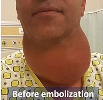
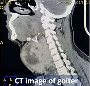
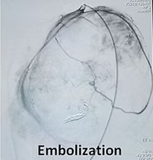
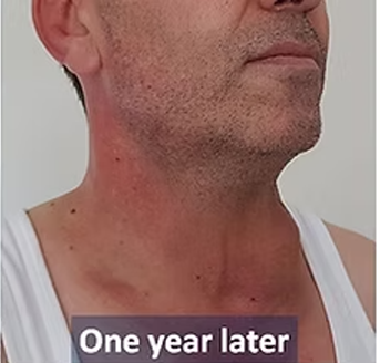
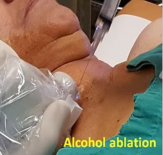
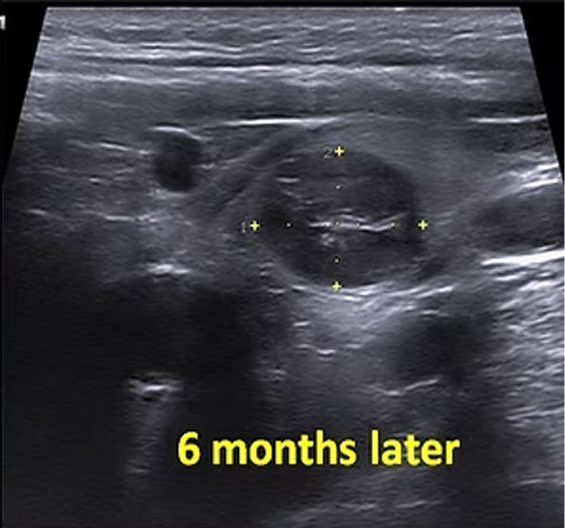
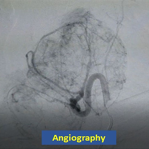
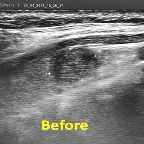
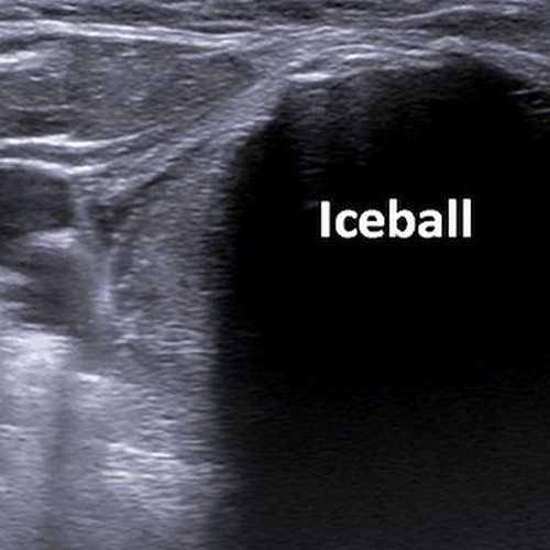
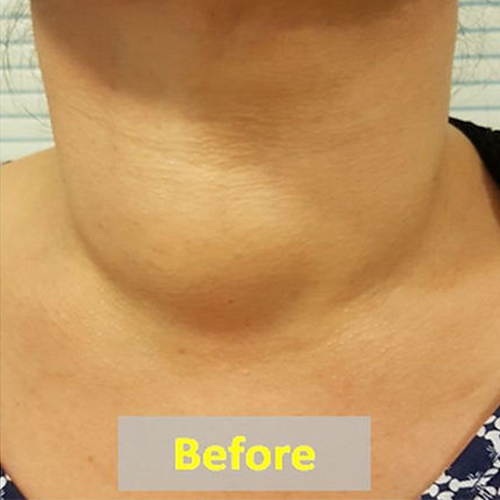
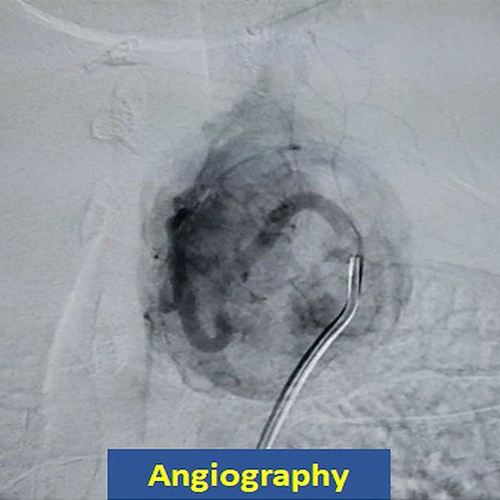
In our 41 year-old patient with a giant goiter, the feeding vessels of the mass were occluded with angiography (embolization). Three and 6 months after the embolization, two sessions of percutaneous ablation were also performed. The goiter mass has shrank continuously over months, and at one year, the patient had almost a normal-looking thyroid gland on ultrasound.

A 72 year old female with a 4cm benign solid nodule in the right thyroid lobe underwent radiofrequency ablation. The patient had hemithyroidectomy 20 years before. Six months after the treatment, the volume of the nodule decreased by 90% and the appearance of the neck returned to normal.

A 45-year-old woman with a giant cystic nodule in the right thyroid lobe underwent ultrasound-guided alcohol (ethanol) ablation. Six months later, the swelling of the patient disappeared, and the ultrasound showed a volume reduction of more than 90%.

A 31 year old male with a 10cm benign solid nodule in the left thyroid lobe underwent embolization. Six months later, the volume of the nodule decreased by 90% and the appearance of the neck was practically normal.

A 59 year old female with a 11 mm papillary carcinoma in the left thyroid lobe underwent percutaneous cryoablation. Nine months later, the nodule was not visible on ultrasound and there was no recurrence during the 1.5 years follow-up.

A 48 year old male with a 7cm benign solid nodule in the left thyroid lobe underwent embolization. Six months later, the volume of the nodule decreased by 80% and the appearance of the neck returned to normal.

In our 32 year-old male patient with a 62x50x47 mm semisolid nodule in the right thyroid lobe, ultrasound-guided alcohol ablation were done 2 times with 3 months interval. One year after the first ablation, a 90% volume reduction was seen on ultrasound.

A 50 year old female with a 13mm incidentally detected papillary carcinoma in the left thyroid lobe underwent percutaneous cryoablation. On ultrasound, a large iceball was formed that completely covered the tumor. Ten months later, the nodule was not visible on ultrasound.

A 65 year old female with a 6.5cm benign solid nodule and multiple small nodules in the left thyroid lobe underwent embolization. Seven months later, the volume of the nodule decreased by 75% and the appearance of the neck returned to normal.

A 70-year-old woman with a giant nodule of 72x53x47 mm in the thyroid istmus region underwent ultrasound-guided microwave ablation. One year later, the swelling of the patient disappeared, and the ultrasound showed a volume reduction of more than 90% in the nodule.

In our patient with a 51x30x37 mm solid nodule in the thyroid, percutaneous laser ablation was done under ultrasound guidance. One year later, the treated nodule has showed 80% volume reduction and the neck returned to normal in appearance.

A 32 year old male with a 6cm benign solid nodule in the left and multiple small nodules in the right thyroid lobe underwent embolization. Six months later, the volume of the large nodule decreased by 85% and the appearance of the neck was grossly normal.

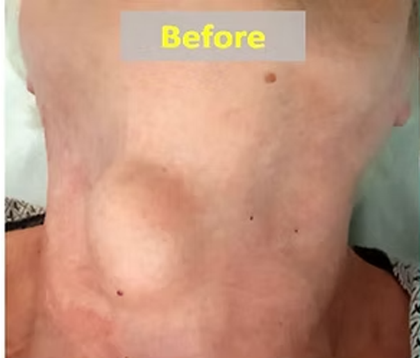
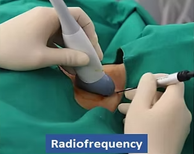
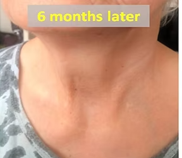
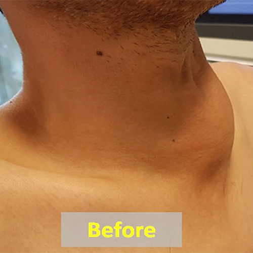
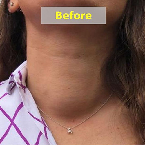
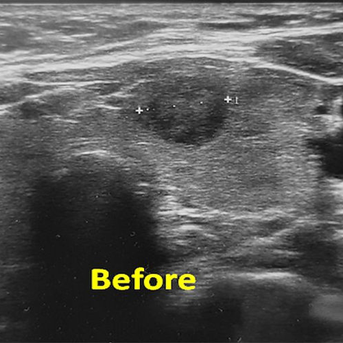
A 34 year old female with a 67 mm solid thyroid nodule underwent radiofrequency ablation. The max diameter of the nodule was 43mm at 3 months. After additional alcohol ablation, the nodule was reduced to 20 mm in size corresponding to more than 90% volume reduction. The patient became symptom-free and the appearance of the neck returned to normal.

In our patient with a large solid nodule of the right thyroid lobe, percutaneous laser ablation was performed under ultrasound guidance. Six months after the treatment, the nodule showed around 70% volumetric shrinkage.

A 26 year old female with a 6cm benign solid nodule and multiple small nodules in the right thyroid lobe underwent embolization. Six months later, the volume of the large nodule decreased by 75% and the appearance of the neck improved markedly.

In our patient with a 38x24x27 mm toxic thyroid nodule, percutaneous laser ablation was performed. One year after the treatment, she became asymptomatic, her thyroid hormone levels returned to normal and the volume of the nodule has reduced by 85%.

A 33 year old male with a 6cm benign solid nodule in the istmus of the thyroid underwent angiographic embolization. Six months later, the volume of the nodule decreased by 80% and the appearance of the neck became grossly normal.

 In our patient with a single, large (53x45x47 mm) solid nodule in the istmus, percutaneous radiofrequency ablation was done. The six months control ultrasound showed over 80% volume reduction of the treated thyroid nodule and the neck returned to normal in appearance.

A 52 year old female with a 12mm incidentally detected papillary microcarcinoma in the left thyroid lobe underwent ultrasound-guided percutaneous microwave ablation. Fifteen months later, the nodule was almost not visible on ultrasound.

In our patient with a $57 \times 50 \times 49$ mm solid nodule in the right lobe, the nodule was treated with percutaneous radiofrequency ablation under ultrasound guidance. The 6 months and 1 year ultrasound examinations demonstrated 60% and 90% volumetric reductions respectively.

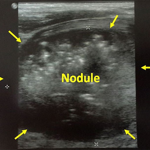
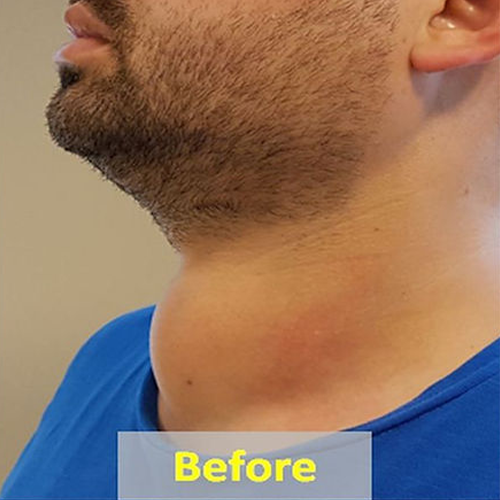
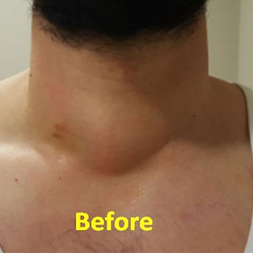
Our 50 year old male patient with a 7cm benign solid nodule in the right thyroid lobe underwent embolization. Five months later, the control CT images show that the diameter of the nodule was reduced from 7cm to 3.7cm, which corresponds to 86% volume reduction. The appearance of the neck also became normal.

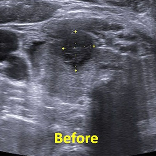
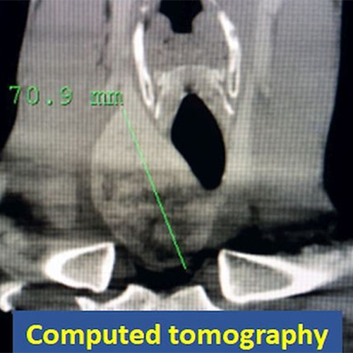
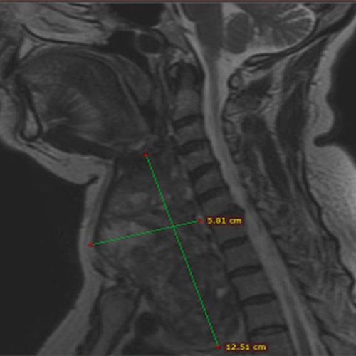
We performed thyroid embolization treatment in our 58-year-old patient with a 12cm giant nodule extending into the chest cavity causing severe dyspnea. A year later, the nodule shrank by 85%, the neck of the patient got slimmer, and shortness of breath disappeared completely. In addition, the mild hyperthyroidism previously present in the patient improved and the thyroid hormones returned to normal.

Our 48 year old male patient with a 9cm benign solid nodule in the right thyroid lobe underwent embolization. Six months after the procedure, the nodule showed 80% volume reduction and the cosmesic appearance of the neck improved markedly.

CONSULTATION FORM

You can get information on our treatments via phone and e-mail as well as by filling and sending the consultation form below. Please send the reports of your thyroid ultrasound, hormones, scintigraphy and biopsy (if available) via e mail (thyroidgoiter@gmail.com) or whats up ( +90-534-551 0 551). Remember to write clearly your e mail address and phone number so that we can return to you as soon as possible.


Please send separately the results of your thyroid ultrasound, hormones, scintigraphy and biopsy (if available)

Please add the reports of your thyroid ultrasound, hormones, scintigraphy and biopsy (if available)